Bacteria Cell Drawing
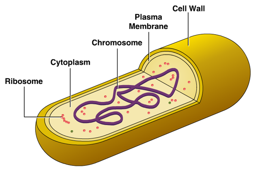
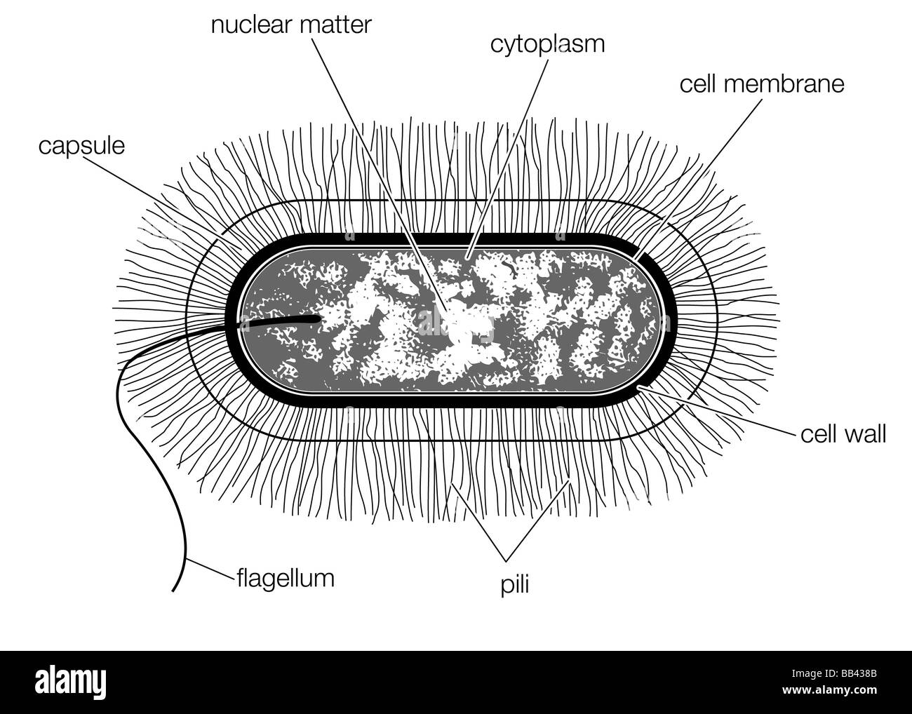
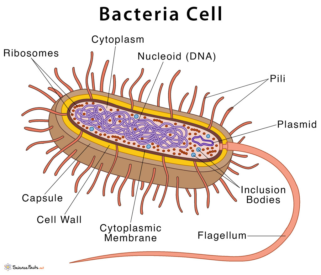
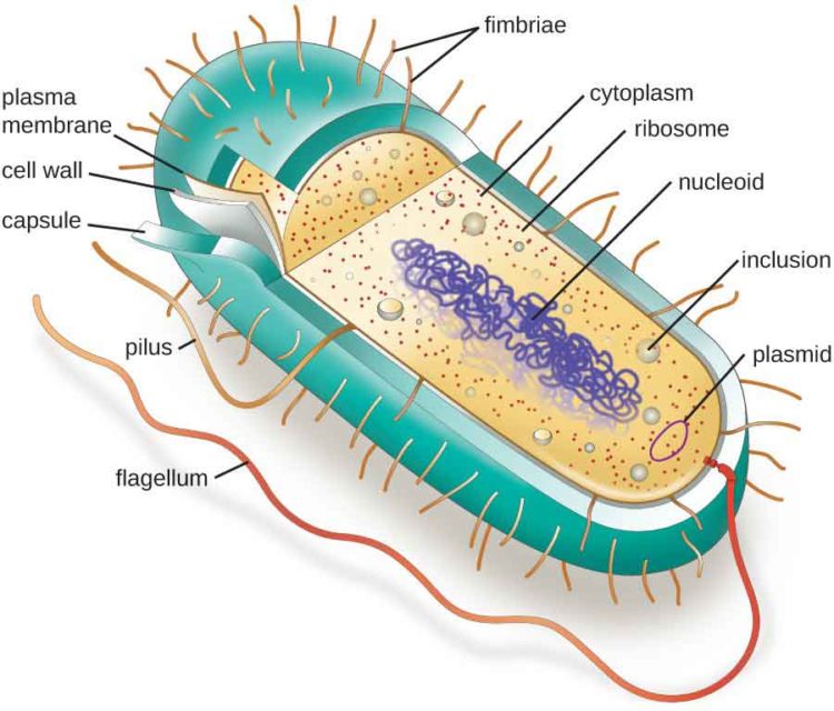
Bacteria Cell Drawing - They come in many shapes and sizes, from minute spheres, cylinders and spiral threads, to flagellated rods, and filamentous chains. There are also cell walls, cytoplasm, and nucleoids (genetic material) are present in the bacterial cell. 38k views 1 year ago #biology #science #ncert. Please don’t forget to subscribe my video. Web the structure of the cell wall of the bacteria determines which dye is visible at the end of the procedure. Web we use to see them via microscope. Web geometric shape features are used to identify the morphological characteristics, namely, flagella and fimbriae or pili of bacterial cells. The cell wall provides an extra layer of protection, helps the cell maintain its shape, and prevents dehydration. Web most bacteria are, however, surrounded by a rigid cell wall made out of peptidoglycan, a polymer composed of linked carbohydrates and small proteins. Web the structure of the bacteria consists of three major parts: The following image is a diagram of a prokaryotic cell; Web we use to see them via microscope. Primary structure of biological macromolecules determines function. The current methods rely on the subjective reading of. Web the bacteria diagram given below represents the structure of a typical bacterial cell with its different parts. In the past century, the discovery and widespread use of antibiotics have somewhat improved the treatment of these infections. Pilus (plural pili) plasma membrane. Web most bacteria are, however, surrounded by a rigid cell wall made out of peptidoglycan, a polymer composed of linked carbohydrates and small proteins. Many structural features are unique to bacteria and. Bacterium) are unicellular prokaryotic microorganisms which divide by binary fission. 38k views 1 year ago #biology #science #ncert. Web most bacteria are, however, surrounded by a rigid cell wall made out of peptidoglycan, a polymer composed of linked carbohydrates and small proteins. The current methods rely on the subjective reading of. Web the bacteria diagram given below represents the structure of a typical bacterial cell with its different parts. By. Many bacteria also have an outermost layer of carbohydrates called the capsule. Web the structure of the bacteria consists of three major parts: Web in this video, we show you how to draw and label a basic bacterial cell. Web geometric shape features are used to identify the morphological characteristics, namely, flagella and fimbriae or pili of bacterial cells. Pilus. Web the bacteria diagram given below represents the structure of a typical bacterial cell with its different parts. They come in many shapes and sizes, from minute spheres, cylinders and spiral threads, to flagellated rods, and filamentous chains. In the past century, the discovery and widespread use of antibiotics have somewhat improved the treatment of these infections. There are also. Web the structure of the cell wall of the bacteria determines which dye is visible at the end of the procedure. Get free printable coloring page of this drawing. Web most bacteria are, however, surrounded by a rigid cell wall made out of peptidoglycan, a polymer composed of linked carbohydrates and small proteins. Bacterium) are unicellular prokaryotic microorganisms which divide. In the past century, the discovery and widespread use of antibiotics have somewhat improved the treatment of these infections. There are also cell walls, cytoplasm, and nucleoids (genetic material) are present in the bacterial cell. Web we use to see them via microscope. The following image is a diagram of a prokaryotic cell; Web most bacteria are, however, surrounded by. The cell wall, plasmid, cytoplasm and flagella are clearly marked in the diagram. Prokaryotic cells do not have a true nucleus that contains their genetic material as eukaryotic cells do. Diplococcus, streptococcus, tetrad, sarcina, and staphylococcus. Web we use to see them via microscope. January 29, 2024 | published on: So in this diagram we try to teach you to draw the parts of bacteria in a easy way. Web the structure of the cell wall of the bacteria determines which dye is visible at the end of the procedure. The cell envelope comprises a rigid cell wall and an underlying cytoplasmic or plasma membrane. Web prokaryotic cell diagram. It. Many bacteria also have an outermost layer of carbohydrates called the capsule. 38k views 1 year ago #biology #science #ncert. Web the structure of a typical bacterial cell with its various components is depicted in the bacteria diagram provided below. Please don’t forget to subscribe my video. The cell wall provides an extra layer of protection, helps the cell maintain. Web bacterial infections have long been a scourge for humanity. Please don’t forget to subscribe my video. In this case, a bacterium. In the past century, the discovery and widespread use of antibiotics have somewhat improved the treatment of these infections. Light and electron microscopes allow us to see inside cells. A typical bacterial cell resembles a plant cell and has a complex membrane, cell walls, cytoplasm, and nucleoids. By following the simple steps, you too can easily draw a perfect bacteria. Little structural detail can be made out in such a small body with an ordinary light microscope. There are also cell walls, cytoplasm, and nucleoids (genetic material) are present. In this article we will discuss about the structure of bacterial cell. Get free printable coloring page of this drawing. Web the bacteria diagram given below represents the structure of a typical bacterial cell with its different parts. The following image is a diagram of a prokaryotic cell; Please don’t forget to subscribe my video. Web bacteria are unicellular organisms with a simple structure. In the past century, the discovery and widespread use of antibiotics have somewhat improved the treatment of these infections. So in this diagram we try to teach you to draw the parts of bacteria in a easy way. This video explains how to draw structure of bacterial. Web the structure of the bacteria consists of three major parts: Web bacterial infections have long been a scourge for humanity. Describe the functions of the structures found in prokaryotic cells. Diplococcus, streptococcus, tetrad, sarcina, and staphylococcus. Procaryotic structural components consist of macromolecules such as dna, rna, proteins, polysaccharides, phospholipids, or some combination thereof. The cell envelope comprises a rigid cell wall and an underlying cytoplasmic or plasma membrane. The cell wall provides an extra layer of protection, helps the cell maintain its shape, and prevents dehydration.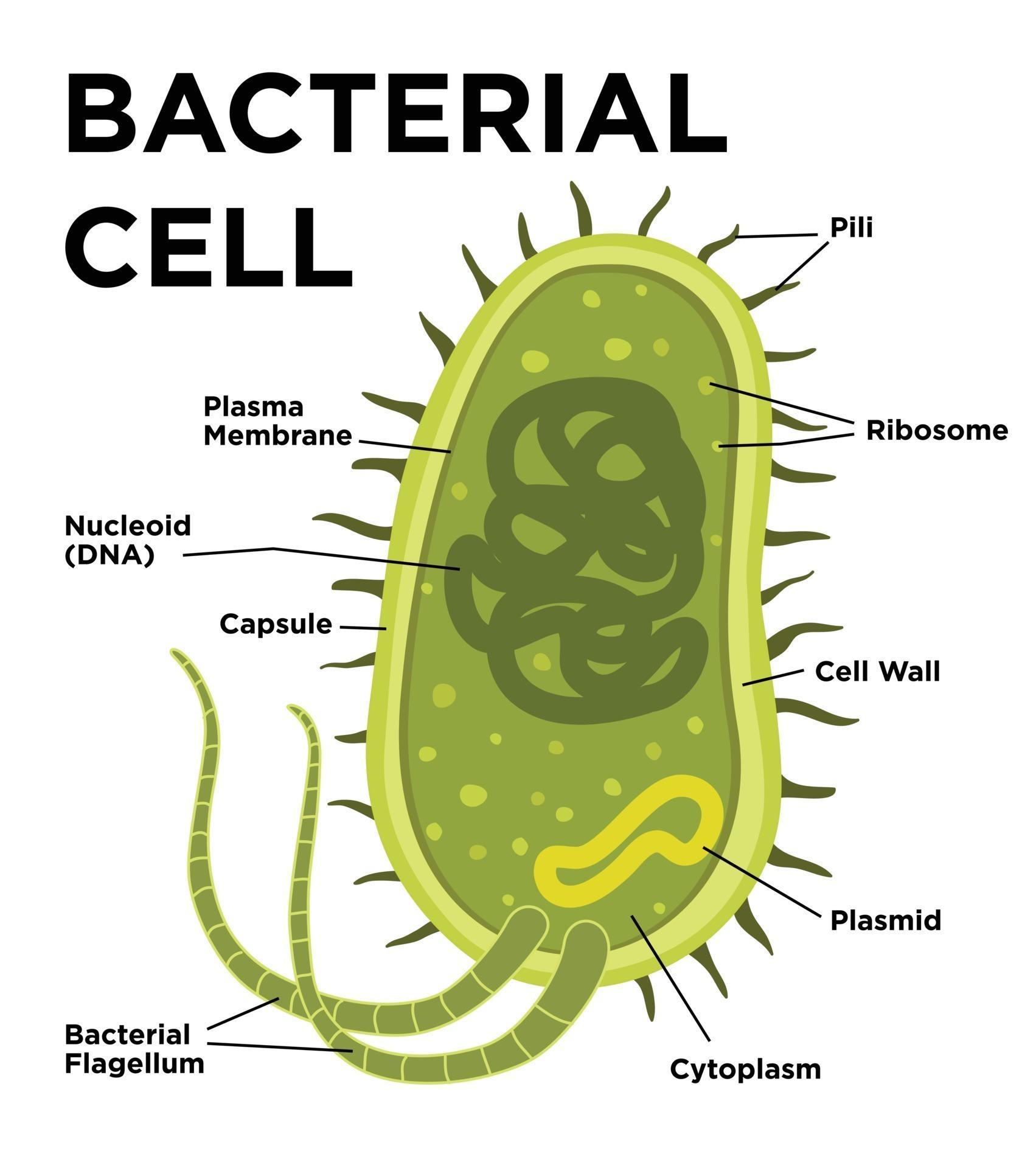
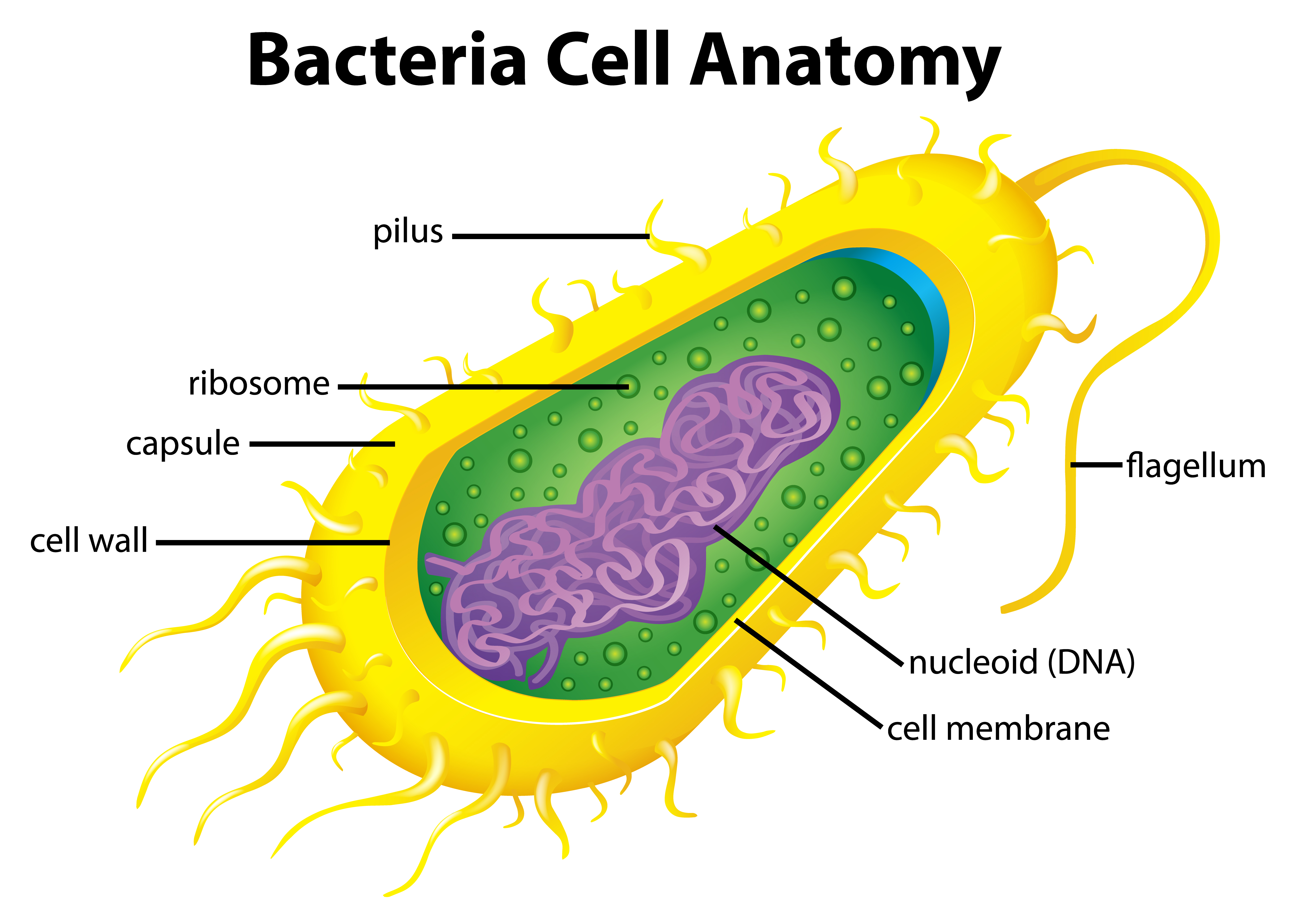
Bacterial cell anatomy in flat style. Vector modern illustration

Bacterial Cell Diagrams 101 Diagrams
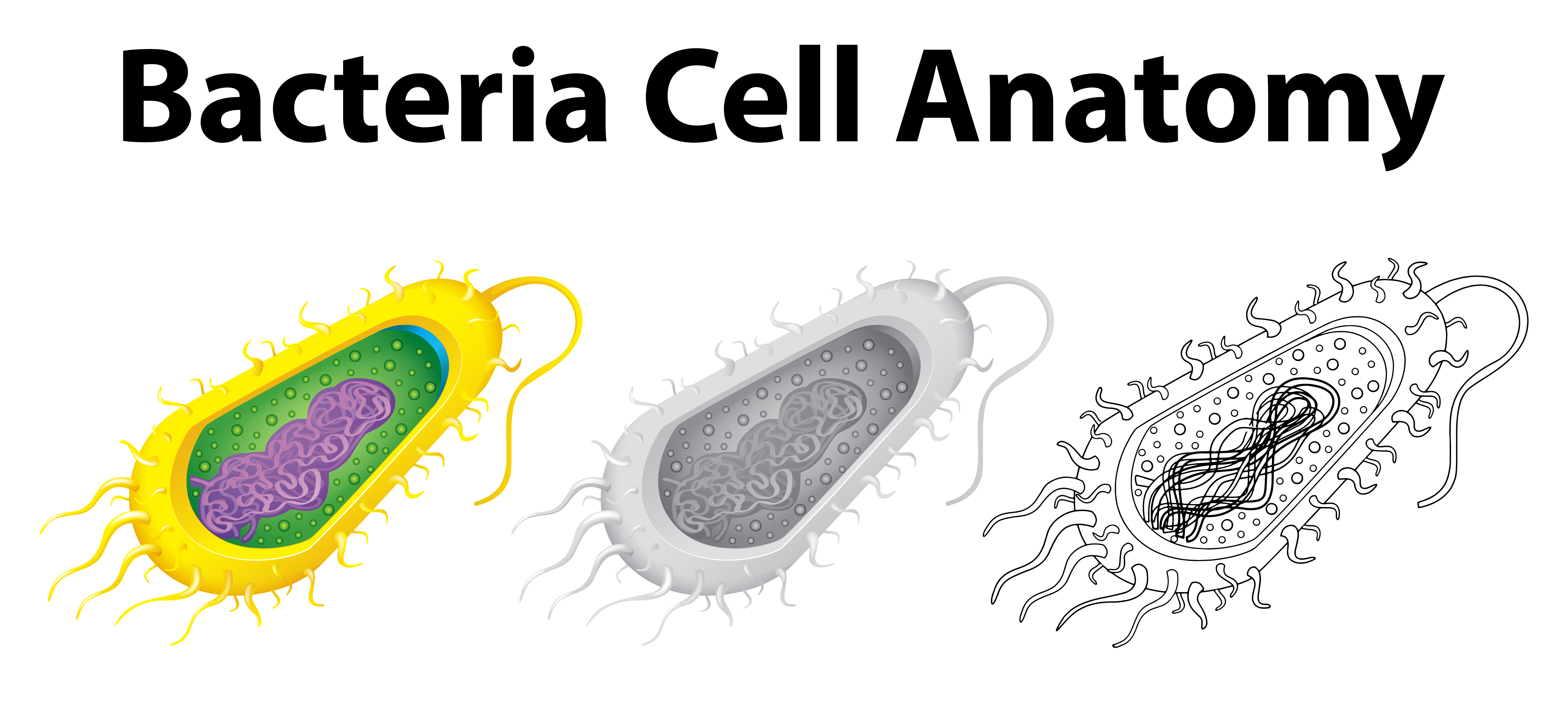
Doodle character for bacteria cell anatomy 431258 Vector Art at Vecteezy

How to Draw Bacteria Really Easy Drawing Tutorial
Bacteria Cell Vector Art, Icons, and Graphics for Free Download
Cellular Structure of Bacteria ZeroInfections
Schematic drawing of the structure of a typical bacterial cell of the
:max_bytes(150000):strip_icc()/bacteria_cell_drawing-5786db0a5f9b5831b54f017c.jpg)
Bacterial Cell Model Labeled
Types Of Bacterial Cells
Bacterial Cell Structure and Function
Web The Structure Of A Typical Bacterial Cell With Its Various Components Is Depicted In The Bacteria Diagram Provided Below.
They Come In Many Shapes And Sizes, From Minute Spheres, Cylinders And Spiral Threads, To Flagellated Rods, And Filamentous Chains.
A Typical Bacterial Cell Resembles A Plant Cell And Has A Complex Membrane, Cell Walls, Cytoplasm, And Nucleoids.
38K Views 1 Year Ago #Biology #Science #Ncert.
Related Post: